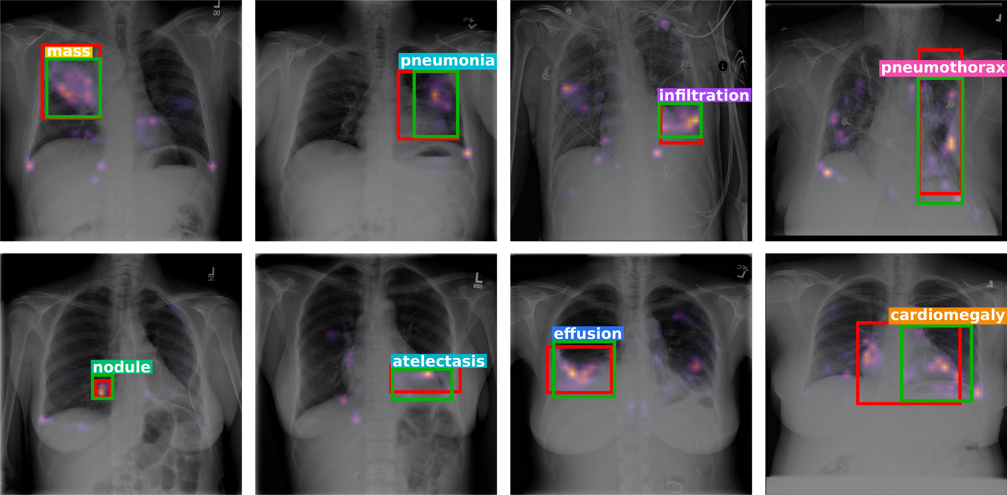
We present ASH-MIL,
Anatomy-Structured Hierarchical MIL for Weakly-Supervised Thoracic Disease Detection in Chest X-rays.
Weakly-supervised thoracic disease detection in chest X-rays (CXR) is challenging due to subtle appearances and complex anatomical overlap, motivating anatomy-aware modeling for improved localization. However, prior anatomy-aware methods typically rely on coarse region proxies or static spatial priors, which may restrict dynamic instance discovery and limit precise localization of small abnormalities.
We propose Anatomy-Structured Hierarchical Multiple Instance Learning (ASH-MIL), a framework that introduces parallel anatomy-structured observation branches (cardiac, pulmonary, and agnostic) combined with hierarchical MIL aggregation. Anatomical priors are injected as soft spatial biases into decoder cross-attention, enabling anatomically grounded evidence maps without disease bounding-box supervision. Instance localization is derived directly from MIL-weighted cross-attention maps without bounding box supervision. Experiments on CXR8 and cross-domain MIMIC-CXR held-out sets demonstrate consistent improvements over prior weakly-supervised and anatomy-aware approaches, particularly under stricter localization criteria.
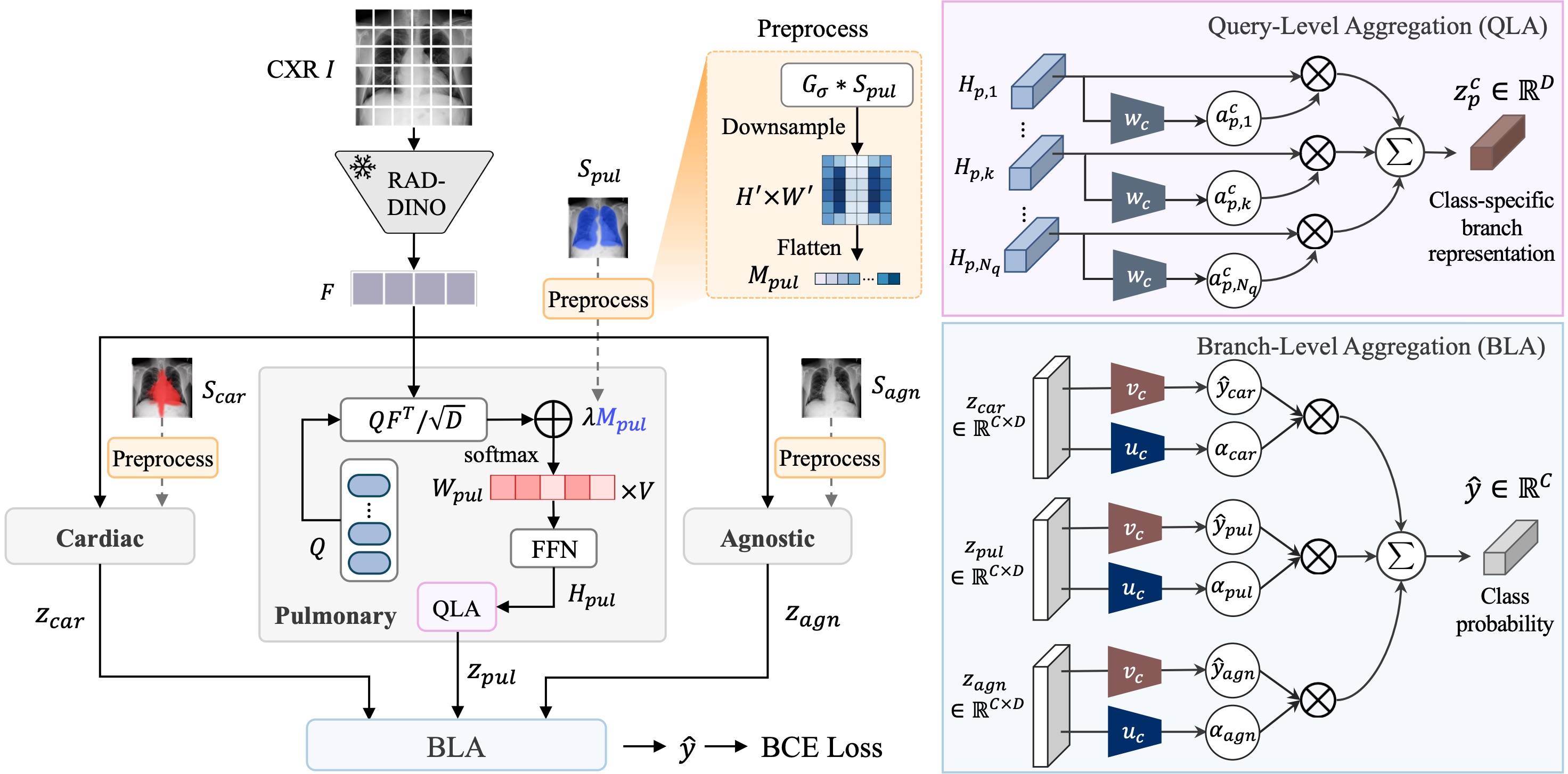
Overall architecture of ASH-MIL. Three parallel branches (cardiac, pulmonary, and anatomy-agnostic) extract complementary evidence from shared image features. Hierarchical MIL combines this evidence across queries and branches for each disease. The pulmonary branch is shown in detail.
ASH-MIL learns where to look for disease evidence and how to combine it, bringing anatomical structure to lesion localization with image-level disease labels.
Anatomy-guided feature encoding. Overlapping structures in chest X-rays can make disease evidence difficult to distinguish. ASH-MIL uses patient-specific organ priors as soft cross-attention biases, guiding learnable queries toward relevant anatomical regions while allowing them to attend beyond organ boundaries. Cardiac and pulmonary branches capture evidence in distinct anatomical contexts, while an anatomy-agnostic branch explores the image without an organ-specific bias.
Hierarchical evidence aggregation. Our hierarchical MIL framework learns which queries and anatomical branches provide useful evidence for each disease.
By combining attention maps with the learned query and branch weights, ASH-MIL localizes suspected lesions and reveals the evidence supporting its predictions, without lesion bounding-box annotations during training.
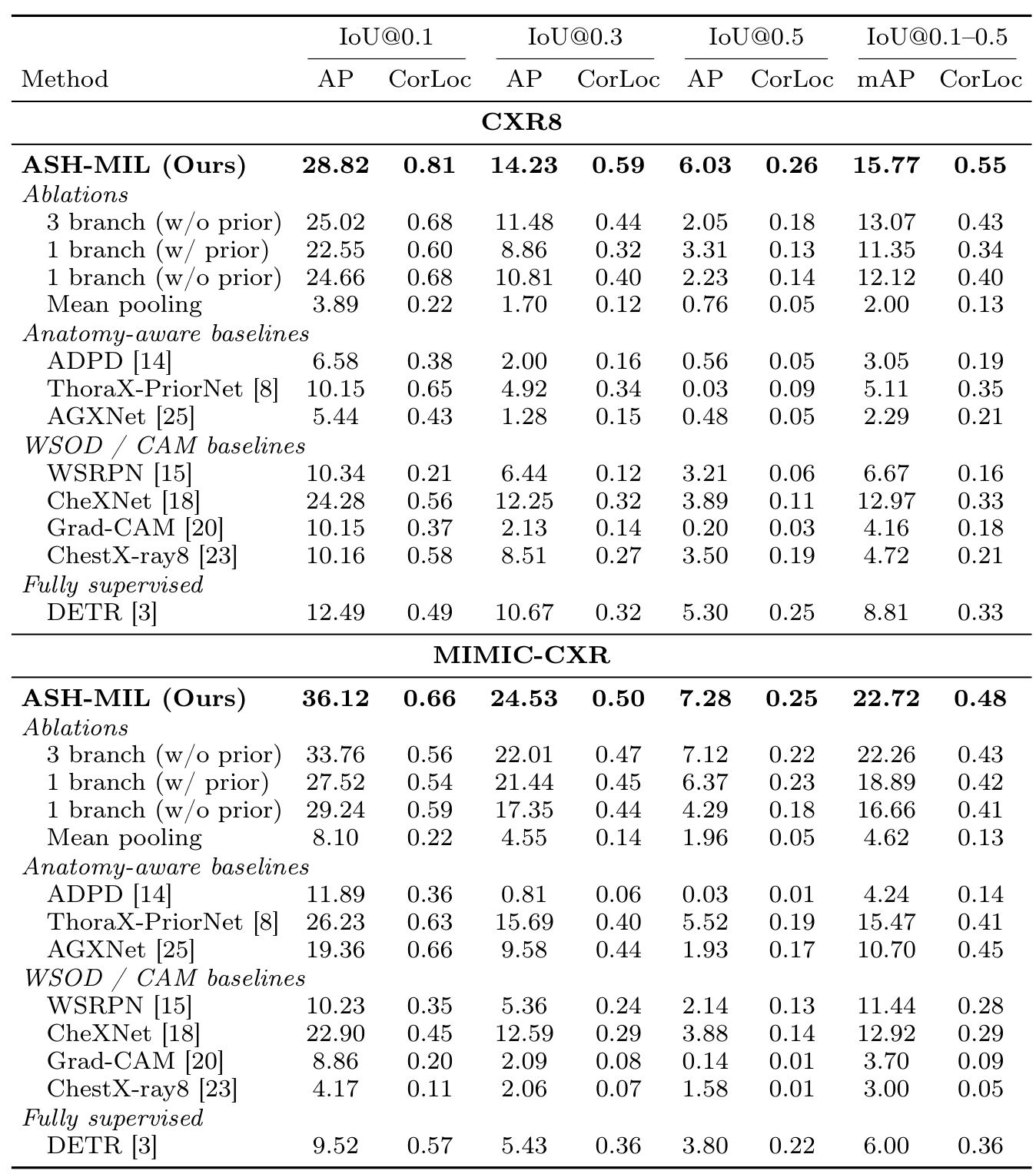
Detection performance and cross-domain generalization. ASH-MIL achieves the highest overall mAP on both CXR8 (in-domain) and MIMIC-CXR (cross-domain). Ablations highlight the complementary benefits of anatomical priors and multi-branch aggregation.
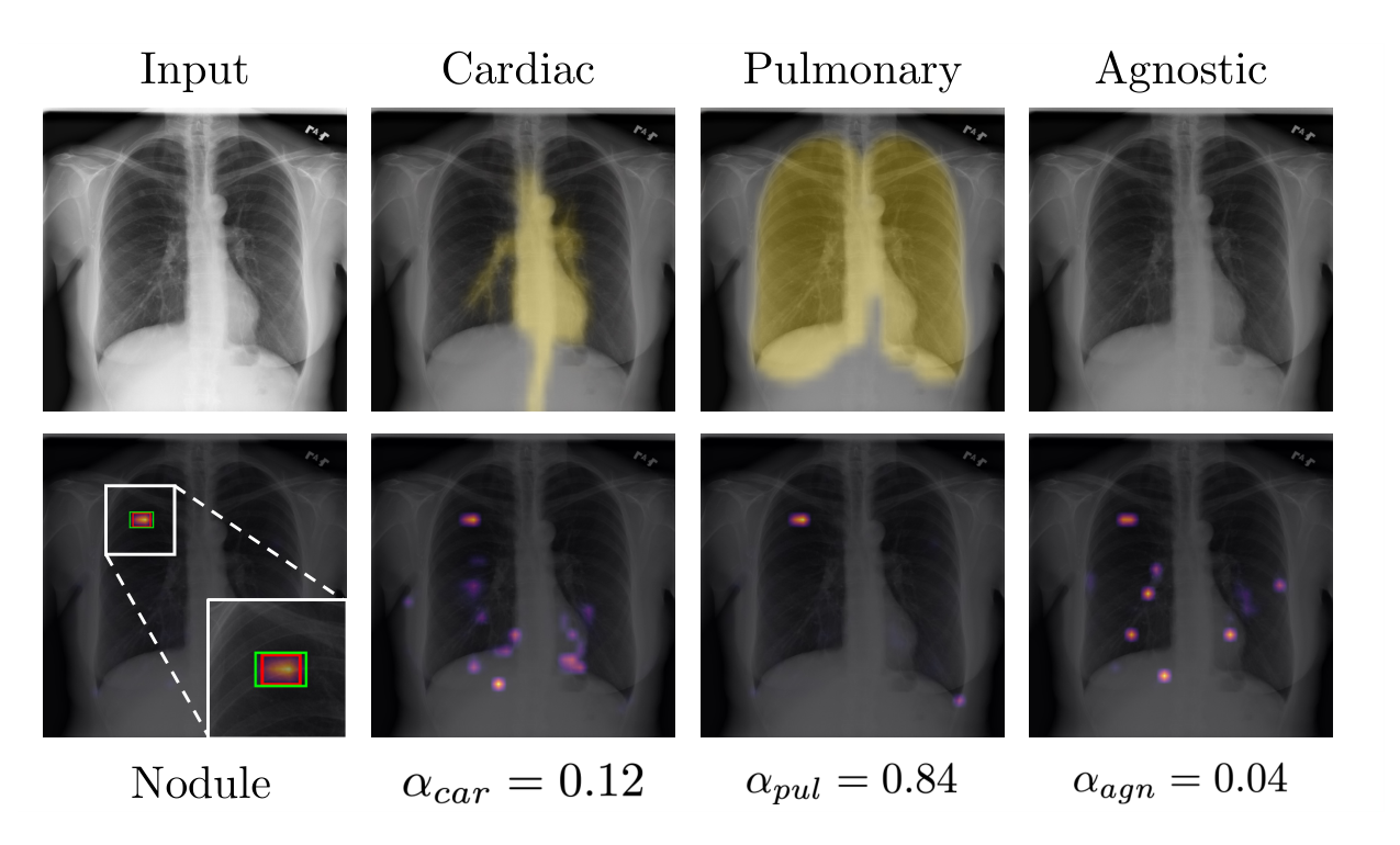
Learning which anatomical evidence matters. For this small nodule within the lung parenchyma, ASH-MIL assigns the greatest weight to the pulmonary branch (0.84), whose evidence map closely aligns with the lesion. Combining the branches with these learned weights produces a compact localization that closely matches the ground truth. Red box: ground truth. Green box: prediction.
@inproceedings{kim2026ashmil,
  title     = {Anatomy-Structured Hierarchical MIL for Weakly-Supervised Thoracic Disease Detection in Chest X-rays},
  author    = {Kim, Jeongin and Ahn, Sohyun and Kang, Seo Young and Sung, Jaeyi and Kim, Soomin and Cho, Sungho and Lee, Rena and Kim, Kwanchang and Noh, Junhyug},
  booktitle = {International Conference on Medical Image Computing and Computer Assisted Intervention (MICCAI)},
  year      = {2026},
}